RAMAN-SNOM MICROSCOPE (alpha300 RS) – OXFORD INSTRUMENTS (WITec)

Origin: Germany

Category:

For the user with challenging experimental requirements, the alpha300 RS facilitates confocal Raman imaging in combination with Scanning Near-field Optical Microscopy for optical imaging with resolution beyond the diffraction limit.

It combines all features of the alpha300 S and alpha300 R and many AFM operation modes. Furthermore the combined Raman-SNOM microscope is ideally suited for high-resolution Raman imaging techniques such as nearfield-Raman imaging.

RAMAN-SNOM MICROSCOPE (alpha300 RS) – OXFORD INSTRUMENTS (WITec)

KEY FEATURES:

  • All features of the alpha300 R (Raman) and the alpha300 S (SNOM) microscope provided in one instrument
  • Excellent combination of high-resolution surface imaging (SNOM) and chemical imaging (Raman)
  • Ideally suited for combined techniques such as near-field Raman imaging
  • Convenient switching between the measurement techniques is realized by a rotation of the objective turret
  • Sample movement between the measurements not necessary

SPECIFICATIONS:

Raman General Operation Modes

SNOM Operation Modes

  • Raman spectral imaging: acquisition of a complete Raman spectra at every image pixel
  • Planar (x-y-direction) and depth scans (z-direction) with manual sample positioning
  • Image stacks: 3D confocal Raman imaging
  • Time series
  • Single point Raman spectrum acquisition
  • Single-point depth profiling
  • Fibre-coupled UHTS spectrometer specifically designed for Raman microsopy and applications with low light intensities
  • Confocal Fluorescence Microscopy
  • Bright Field Microscopy
  • Scanning Near-field Optical Microscopy (SNOM) modes: bottom-up and top-down mode, collection mode
  • Confocal Microscopy (CM) modes:
  1. Transmission
  2. Reflection
  3. Fluorescence (optional)
  4. SNOM-AFM combinations: AFM operation modes of alpha300 A included or optional available
  5. Acquisition of force-distance curves and light-distance curves
  6. Fixed-bottom illumination
  7. Total internal reflection illumination (optional)


Basic Microscope Features

Raman Optional/Upgradable Operation modes

  • Research grade optical microscope with 6x objective turret
  • Video system: video CCD camera
  • High sensitivity b/w video camera to view sample and SNOM/AFM tip in transmission
  • LED white-light source for Köhler illumination of AFM tip and sample
  • Manual sample positioning in x- and y-direction
  • Microscope base with active vibration isolation system
  • Fibre coupling
  • Additional lasers, several wavelenghts eligible
  • Additional UHTS spectrometers (UV, VIS, NIR)
  • Automated, motorized sample positioning and measuring with piezo-driven scan stages
  • Automated confocal Raman imaging
  • Automated multi-area and multi-point measurements
  • Ultrafast Raman imaging (1300 spectra per second) optionally available
  • Upgradable for epi-fluorescence applications
  • Adapter for higher samples
  • TrueSurface for Raman depth profiling
  • Autofocus


AFM Operation Modes

Computer Interface

  • Contact Mode
  • Lateral Force Mode
  • Others optional
  • WITec software suite for instrument and measurement control, data evaluation and processing

APPLICATION EXAMPLES:

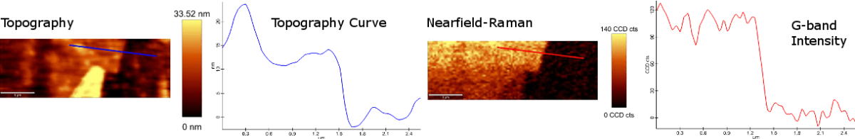
Left: Topography image of exfoliated graphene simultaneously determined during the nearfield-Raman measurement with corresponding topography curve measured along the blue line.

Right: Nearfield-Raman image of the same sample area of the G-band intensity with corresponding intensity graph measured along the red line.

PRODUCT VIEWED

No recently viewed products